Treatments
Sonography & 2d Echo
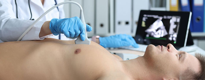
Sonography & 2D Echo are both essential diagnostic tools used in medical imaging to assess various aspects of health, especially for cardiovascular and obstetric care. They use ultrasound technology to create images of internal structures, providing valuable information for diagnosing and monitoring a wide range of conditions.
Sonography (Ultrasound Imaging)
Sonography, also known as ultrasound imaging, uses high-frequency sound waves to create images of the organs and structures inside the body. It is non-invasive, safe, and commonly used to evaluate the following:
Obstetric Care
- Pregnancy Monitoring: Sonography is widely used to monitor fetal growth, development, and position during pregnancy. It can detect abnormalities, confirm gestational age, and assess the health of the baby.
- Early Detection of Complications: It helps in identifying conditions such as ectopic pregnancies, fetal heart defects, and multiple pregnancies (twins, triplets).
Abdominal Imaging
- Liver, Kidneys, and Gallbladder: Sonography is used to evaluate the size, structure, and any abnormal growths or diseases in organs like the liver, kidneys, pancreas, and gallbladder.
- Detection of Cysts or Tumors: It can identify the presence of cysts, tumors, or stones in organs.
Cardiovascular
- Assessing Blood Flow: Doppler ultrasound, a type of sonography, evaluates blood flow in arteries and veins, useful in diagnosing conditions like deep vein thrombosis (DVT), arterial blockages, and peripheral artery disease.
Musculoskeletal Imaging
- Joint and Soft Tissue: Used to assess soft tissue injuries, joint inflammation, tendon injuries, and musculoskeletal conditions.
2D Echocardiography (2D Echo)
2D Echocardiography or 2D Echo is a specialized form of ultrasound used to visualize the heart's structure and function in two dimensions. It helps in diagnosing and monitoring various cardiovascular conditions. The process uses high-frequency sound waves to create images of the heart's chambers, valves, and blood vessels.
- Heart Function Assessment: It evaluates the heart's ability to pump blood, which is crucial in conditions like heart failure or cardiomyopathy.
- Valvular Heart Disease: 2D echo is instrumental in detecting heart valve problems like stenosis (narrowing) or regurgitation (leakage), which can affect blood flow.
- Congenital Heart Defects: It helps diagnose structural heart issues present from birth, such as septal defects (holes in the heart wall) or valve malformations.
- Pericardial Effusion: This test can detect fluid buildup around the heart (pericardial effusion), a potentially dangerous condition that can affect heart function.
- Aortic Aneurysm: It can identify an enlarged aorta or aneurysm, which can be life-threatening if not detected early.
- Monitoring of Previous Conditions: Post-surgical monitoring: 2D Echo is also used to monitor patients who have undergone heart surgery, such as valve replacements or bypass procedures.